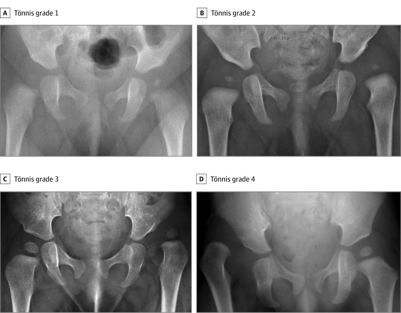
Figure 1. Tönnis Classification.
A, Grade 1: the center of the femoral head lies medial to the Perkin line (a vertical line drawn from the most lateral point of the acetabulum). Radiograph is of an infant girl with a dislocatable right hip discovered at routine examination. B, Grade 2: the center of the left femoral head lies lateral to the Perkin line. Radiograph is of an infant girl referred because of decreased hip abduction at routine examination. C, Grade 3: the center of the left femoral head lies at the level of the lateral aspect of the acetabulum. Radiograph is of an infant girl referred because of limited abduction. The mother had noted shortening of the leg. D, Grade 4: the center of the right femoral head lies above the lateral aspect of the acetabulum. Radiograph is of a male child referred because of leg length discrepancy. The parents had sought medical attention for some time before the diagnosis was made.

