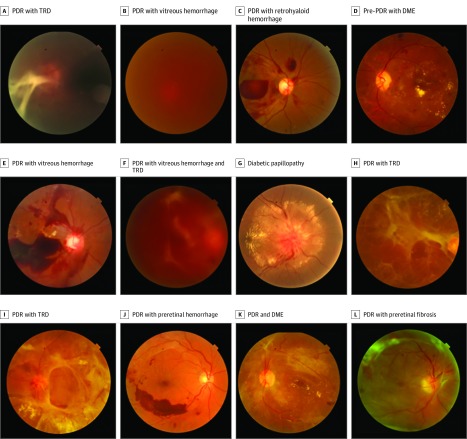
Figure 1. Examples of Disc-Centered and Macula-Centered Screening Photographs.
A, Proliferative diabetic retinopathy (PDR) with tractional retinal detachment (TRD) (retinopathy [R]3). B, PDR with vitreous hemorrhage (R3). C, PDR with retrohyaloid hemorrhage (R3). D, Pre-PDR with diabetic macular edema (DME) (R2 maculopathy [M]1). E, PDR with vitreous hemorrhage (R3). F, PDR with vitreous hemorrhage and TRD (R3). G, Diabetic papillopathy. H, PDR with TRD (R3). I, PDR with TRD (R3). J, PDR with preretinal hemorrhage (R3). K, PDR and DME (R3M1). L, PDR with preretinal fibrosis (R3).

