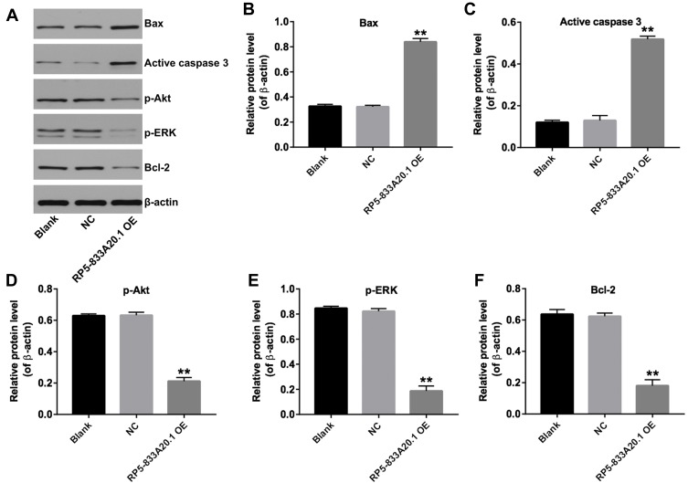
Figure 5.
Overexpression of RP5-833A20.1 induced apoptosis of HCC cells via inhibiting Akt/ERK pathway. (A) Huh7 cells were infected with NC or lenti-RP5‑833A20.1 for 72hrs. Expression levels of Bax, active caspase 3, p-Akt, p-ERK and Bcl-2 in Huh-7 cells were detected with Western blotting. β-actin was used as an internal control. (B–F) The relative expressions of Bax, active caspase 3, p-Akt, p-ERK and Bcl-2 in Huh-7 cells were quantified via normalization to β-actin. **P<0.01 vs NC group.

