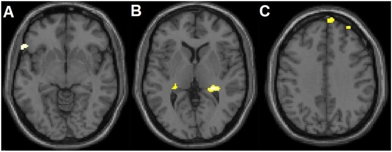
Figure 3. Additional Grey Matter Volume Differences between Adolescents and Young Adults with Bipolar Disorder with History of Suicide Attempts, Compared to Without Suicide Attempts.
The structural magnetic resonance T1 axial-oblique images display additional regions where grey matter volume was significantly lower in attempters than non-attempters only within the bipolar disorder group, including A) left ventral lateral prefrontal cortex, B) bilateral hippocampus and C) right dorsomedial and dorsolateral prefrontal cortex (p<0.005 uncorrected and spatial extent of 20 contiguous voxels). The right side of the axial-oblique images is the right side of the brain.

