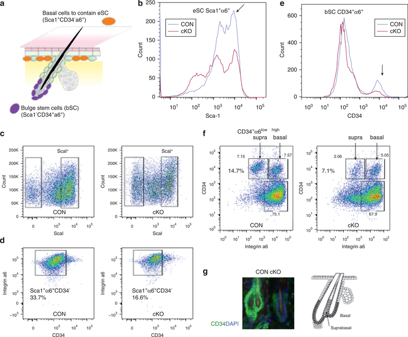
Figure 2. VDR deficiency decreased self-renewal potentials for epidermal SC.
FACS analyses for basal cells containing eSCs (Sca1+α6+CD34−) and bSCs (Sca1−α6+CD34+) in VDR cKO and CON are shown. (a) The scheme to show the location and expression markers for bSCs and eSCs. (b, e) Representative histograms for Sca1 (b) and (e) for CD34 to compare CON (blue) and cKO (red) are shown. (c, d) The sorting profiles and number of basal cells containing eSCs (Sca1+α6+CD34-). (f) The profiles and number for suprabasal bSCs (Sca1−α6lowCD34+) and basal bSCs (Sca1+α6highCD34−). (g) Immunostaining of CD34 (CD34 green, DAPI blue) on HFs (3-month) with a diagram to show locations of basal and suprabasal bSCs. These results were reproduced in two experiments and representative data are shown. α6, integrin α6; bSC, hair follicle bulge stem cell; cKO, conditional knockout; CON, control; eSC, interfollicular epidermal stem cell; VDR, vitamin D receptor.

