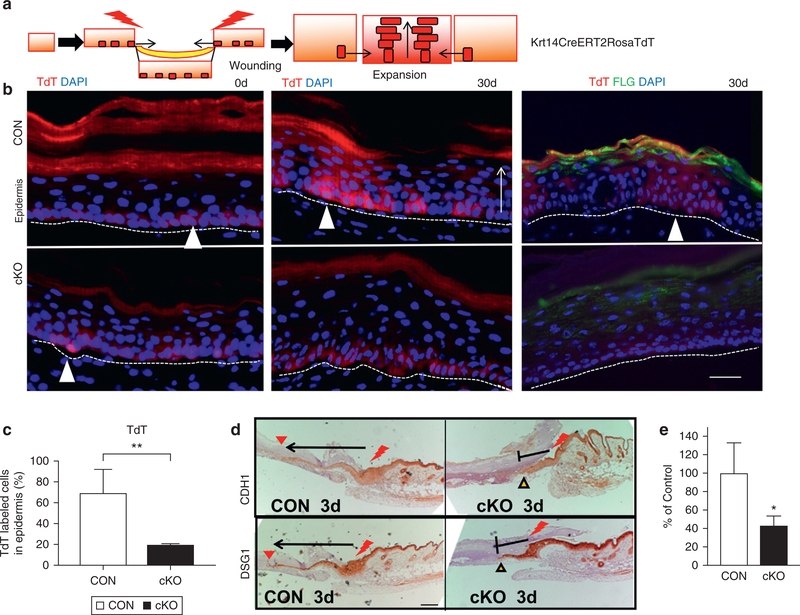
Figure 4. VDR ablation impairs SC-driven epidermal regeneration during wound re-epithelialization.

(a) The strategy for lineage tracing using Krt14CreERT2RosaTdT. (b) Representative images of initial skin biopsy (0 days) (left panels) and after 30 days of wound healing (middle panels), TdT-labeled SCs migrated to form clones in CON (white triangles, upper panels), but not in cKO (bottom panels). Differentiation was verified by FLG (right two panels) (TdT red; FLG green; DAPI blue). Scale bar = 25 μm (c) Quantification of the TdT-labeled cells in the epidermis by Bioquant (**P < 0.05). (d) Migration of epithelial tongues at 3 days, in which wounding edge is shown by red bolt, and the extent of the epithelial tongue is shown by arrows. Scale bar = 100 μm. (e) Quantitated migration at 3 days (n = 3, P < 0.05). cKO, conditional knockout CON, control; SC, stem cell; TdT, TdTomato; VDR, vitamin D receptor.
