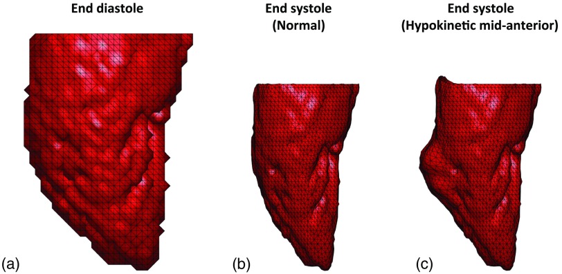
Fig. 3.
End-diastolic and analytically derived end-systolic LV poses. (a) End-diastolic mesh derived from human LV clinical CT data. (b) Analytically derived end-systolic pose with normal function. (c) Analytically derived end-systolic pose exhibiting regional hypokinesia of 1/2 an AHA segment (14 mm) as core diameter at the midanterior segment of the LV with normal strain values in all other segments. All three views are of the LV lateral wall with the anterior wall to the left and the inferior wall to the right.

