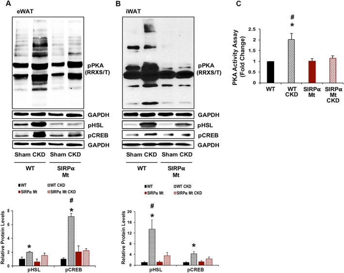
Figure 4.

PKA activity and its mediators are suppressed in SIRPα Mt Mice with CKD. (A–B) eWAT and iWAT protein lysates were immunoblotted to detect phosphorylated protein kinase A [pPKA (RRXS/T)], pHSL, pCREB, and GAPDH (top panel); and the relative densities to the level of GAPDH are shown (bottom panel). (C) PKA activity assay was determined in eWAT (A–C: n = 4–6 mice/group). Values are expressed as mean ± SEM; *P < 0.05, sham vs. CKD and #P < 0.05, WT vs. SIRPα Mt mice.
