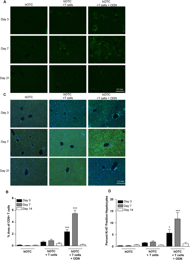
Fig. 3.
T-cell infiltration and hepatocyte proliferation. Mice received i.v. injections of the vector and, 14 days later, OT-1 CD8+ T cells were transferred concurrent with an administration of 40 μg CpG ODN. Mice were sacrificed on days three, seven, and 14 following adoptive transfer. (A) We stained liver sections for CD8, and (B) we determined the percent area covered by infiltrating T cells using Image J. (C) We stained liver sections with the Ki-67 proliferation marker, and (D) calculated percent positive hepatocytes. For image analysis experiments, we analyzed five images per mouse. N = 4 Statistical analysis by ANOVA with a Bonferroni posttest to day appropriate vector only control. . *P < 0.05, ***P < 0.0001. Error Bars = SEM.

