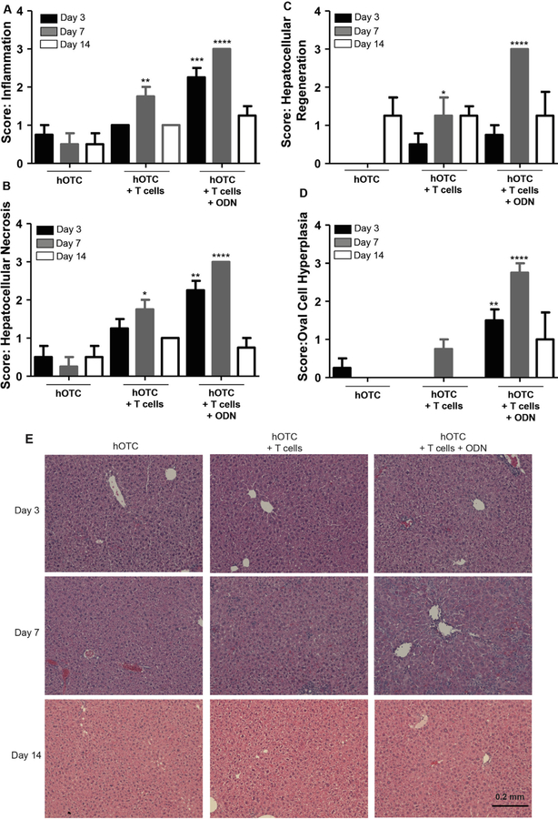
Fig. 4.
Liver pathology following adoptive transfer. Mice received i.v. injections of the vector and, 14 days later, OT-1 CD8+ T cells were transferred concurrent with an administration of 40 μg CpG ODN. Mice were sacrificed on days three, seven, and 14 following adoptive transfer. We stained liver sections for H&E and scored for extent of (A) inflammation, (B) hepatocellular necrosis, (C) hepatocellular regeneration, and (D) oval-cell hyperplasia. (E) The histopathologic images depicted represent general trends for each dose group. Statistical analysis by student t test. *P < 0.05, **P < 0.001, ***P < 0.0001. ****P < 0.00001. Error Bars = SEM.

