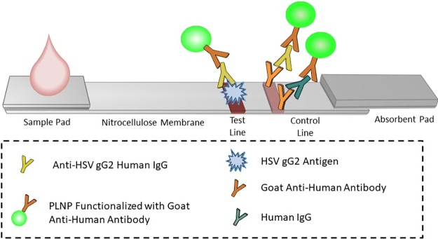
Fig 1. HSV-2 PLNP LFA schematic.
A sample is diluted and then mixed with PLNPs functionalized with goat anti-human IgGs to form human IgG-PLNP complexes that were dispensed onto the sample pad of the LFA strip. The anti-HSV human IgG PLNP complexes migrated up the membrane and were captured by recombinant HSV gG2 immobilized at the test line. The remaining uncaptured human IgG-PLNP complexes, whether specific to HSV2 gG2 or not, continued further up the strip until they were captured by goat anti-human IgGs immobilized at the control line.

