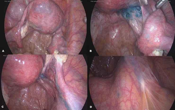
Fig 2. Laparoscopic images during a tubal dye test.
The uterus showed a transient scattered blue color and unilateral right tubal permeability was demonstrated (A). A deep blue color of the endometriosis nodule of the right uterosacral ligament appeared after a few seconds (B) along with a progressive staining of lymphatic vessels of the infundibulopelvis ligament (C). A blue spot was seen where lymph nodes of the internal iliac vein lie (D).

