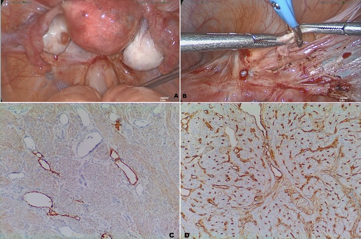
Fig 4. Laparoscopic and histologic images.
Clustered areas of the uterus were permeated by the methylene blue during a dye test that showed bilateral permeability of the tubes (A). After opening the peritoneum of the anterior broad ligament, a blue staining of the connective tissue beneath the round ligament (*) was found (B). In this case, a deep biopsy of the uterine wall where the color changed was taken and adenomyosis with ectatic lymphatic vessels was demonstrated. Deparaffinized 4-μm sections were immunostained with antibodies against CD34 and podoplanin (C and D, respectively) to confirm that the ectatic vessels were lymphatics.

