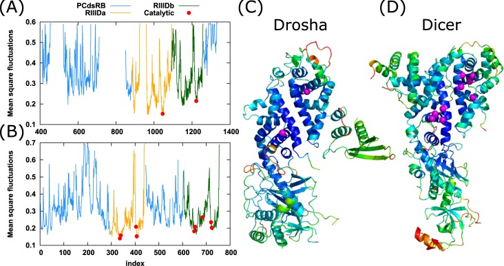
Fig 4. GNM mean-square fluctuations profile.
Distribution of mean-square fluctuations (ordinate) as a function of residue index (abscissa) for (A) Drosha and (B) Dicer. Peaks represent the most mobile residues and minima the low mobility regions. Fragmented areas in the curve are UNK or missing residues in the PDB files. Curve colors are as follow: non catalytic domains (light blue; PCdsRB; platform, connector, dsRBD, bridging domain), RIIIDa (orange), RIIIDb (green). Catalytic residues are marked by filled red circles. (C) Drosha and (D) Dicer ribbon diagram is colored according to their MSF values. Warm and cold colors (red and blue scales, respectively) indicate most mobile and constrained regions, respectively. PCdsR designate: platform, connector, and dsRBD domains.

