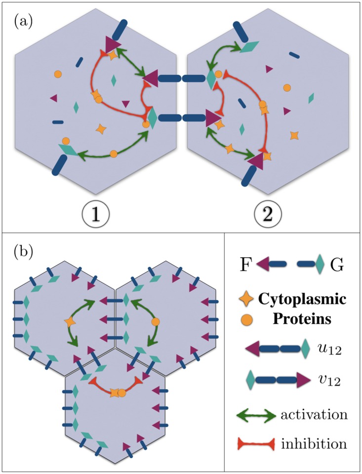
Fig 1.

(a) A schematic of intracellular and intercellular mechanisms in adjacent cells. Each cell contains a pool of membrane and cytoplasmic proteins. Membrane proteins Fz (red triangles ▲) and Vang (green diamonds ♦) bind to the transmembrane proteins Fmi (dark blue bars), and form cross-junctional complexes F—G. Two opposite complexes localized on the cell-cell junction are shown in the figure. Nonlocal interactions, illustrated by orange stars ★ and solid disks ●, are mediated by diffusing cytoplasmic proteins that couple the membrane-bound proteins. Star-shaped proteins bind to the red triangles (Fz), get modified and released back to the cytoplasm, to either upregulate the formation of same-polarity complexes, or downregulate the opposite ones; similarly for the orange disks and green diamonds (Vang). While the upregulating interactions between similar proteins, i.e. Fz ↔ Fz, or Vang ↔ Vang, are transmitted by their associated cytoplasmic proteins, stars and disks, respectively, both types of these cytoplasmic proteins participate in the downregulating of the opposite complexes. In order to keep the picture clear, only a few of the pairwise interactions are drawn. (b) shows the protein distributions in a polarized state, where the segregation of F and G is accomplished in each cell through cytoplasmic interactions.
