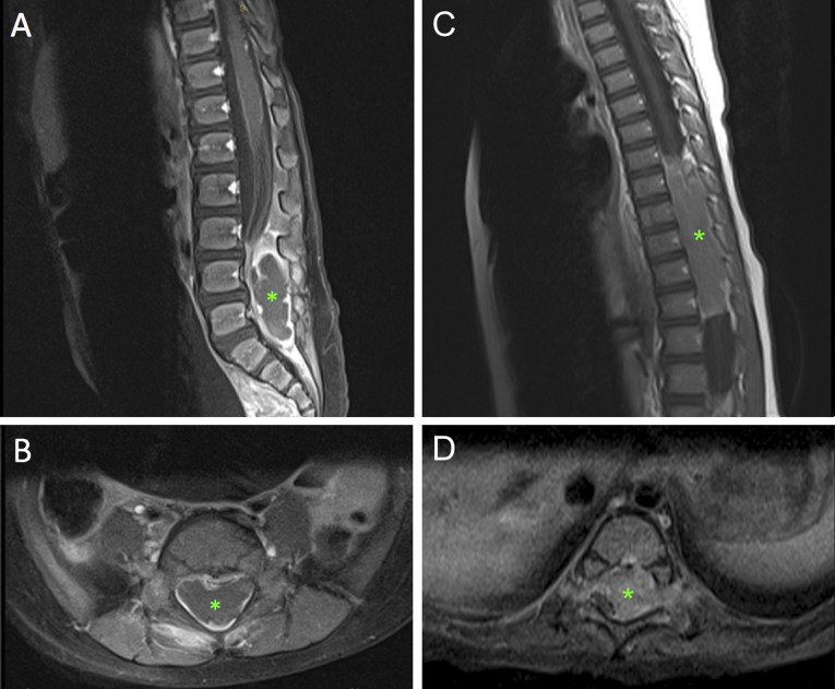
Figure 1.
(A and B) Case 1 T1-weighted postcontrast preoperative sagittal and axial spinal MRI. Asterisk indicates a 5.3 x 1.5 x 2.3-cm epidural mass extending from L3 to S1 with foraminal involvement. The mass has a rim-enhancing component causing notable ventral displacement of the cauda equine. (C and D) Case 2 T1-weighted postcontrast preoperative sagittal and axial spinal MRI. Asterisk indicates a mass extending from T7 to 12 with dorsal and right-sided displacement of the spinal cord with bilateral foraminal extension.

