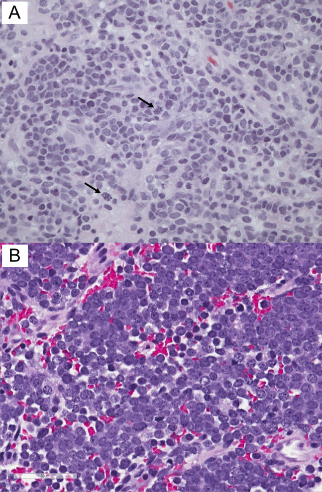
Figure 3.
(A) Case 1 histology shows undifferentiated small blue cells consisting of round to oval hyperchromatic nuclei, no nucleoli, and scant cytoplasm. Cells were arranged in sheets with strands of collagenous stroma, forming vaguely lobular architecture. Arrows indicate mitoses. (B) Case 2 histology shows sheets and small clusters of small round blue cells with background hemorrhage and necrosis. The tumor diffusely infiltrates the surrounding fibrous tissue (hematoxylin and eosin stain, ×400).

