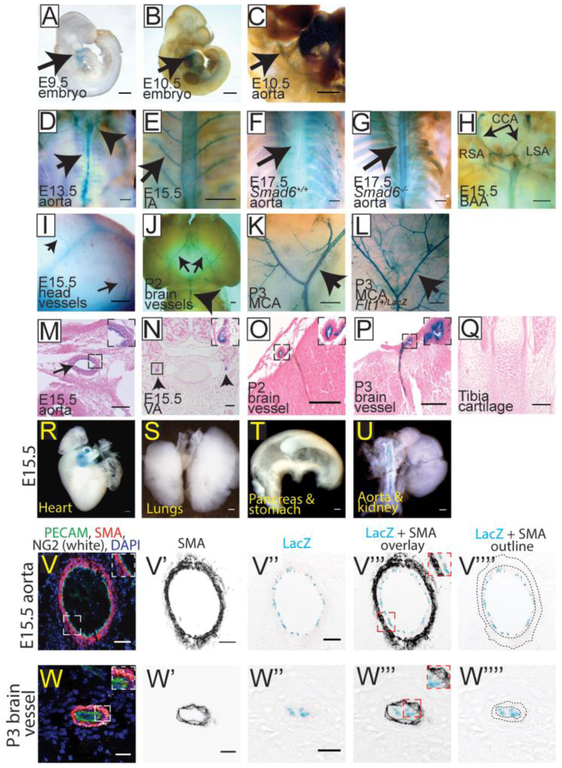
Figure 2: SMAD6 is Selectively Expressed in a Subset of Vessels Developmentally.
A-L Whole embryos stained for LacZ expression (blue). M-Q, cross sections of embryos stained for LacZ expression, counterstained with eosin (pink). A, E9.5 embryo with LacZ expression in the heart and outflow tract (arrow, heart). B-C, E10.5 embryo showing LacZ expression in heart and dorsal aorta (DA) (arrows, DA). C) Higher magnification of DA staining in (B). D-G, Body cavities of E13.5 Smad6−/− (D), E15.5 Smad6−/− (E), and E17.5 Smad6+/+ (F) and Smad6−/− (G) embryos with all organs removed to show LacZ expression in aorta (black arrows in D, G) and intercostal arteries (IA) (arrow in E), compared to only background in Smad6+/+ embryo (F) (Arrowhead in D, branchial arch arteries). H, E15.5 Smad6−/− branchial arch arteries (BAA) positive for LacZ expression. CCA, common carotid arteries; LSA, left subclavian artery; RSA, right subclavian artery. I, Blood vessels expressing LacZ in E15.5 Smad6−/− embryo head (arrows, vessels). J-K, Postnatal (P2-3) Smad6−/− brains showing arterial LacZ expression. MCA, Middle cerebral arteries; BA, basilar artery (arrows J-K, MCA; arrowhead J, BA). L, Flt1LacZ P3 brain showing arterial, venous, and capillary LacZ expression; arrow to MCA. M, Cross-section showing LacZ expression in aorta of E15.5 Smad6−/− embryo (arrow, aorta); inset, higher magnification of aorta. N, E15.5 Smad6−/− embryo cross-section with staining in smaller vertebral arteries (arrows, vertebral arteries); inset, higher magnification of left vertebral artery. O-P, Cross-sections of postnatal (P2-3) brains; insets, higher magnification of indicated vessel. Q, Cross-section of E15.5 tibia cartilage showing no detectable LacZ expression. R-U, Whole mount of indicated E15.5 organs stained for LacZ expression. LacZ expression is detected in the outflow tract of the heart (R), but not in non-vascular tissue of lungs (S), stomach and pancreas (T), or kidney (U, note LacZ expression in aorta connected to kidney). V-W””, E15.5 Smad6−/− aorta (V-V””) and P3 brain (W-W””) serial cross-sections stained with α–smooth muscle actin (SMA, red), PECAM (green), NG2 (white) and DAPI (blue) in V, W. V’, W’, SMA channel alone. V”, W”, LacZ reporter expression (blue) in same cross-section as in V’ and W. V”’-W”’, proportional overlay of SMA and LacZ expression patterns (compare insets in V, W to insets in V”’, W”’). V””, W””, SMA layer outline (dotted lines) over LacZ expression image, showing no apparent overlap between SMA and LacZ (SMAD6) expression. Scale bars: A-I, 500μm; J-L, R-U, 1,000μm; M-Q, 100μm; V-V””, 50μm; W-W””, 20μm.

