Figure 3.
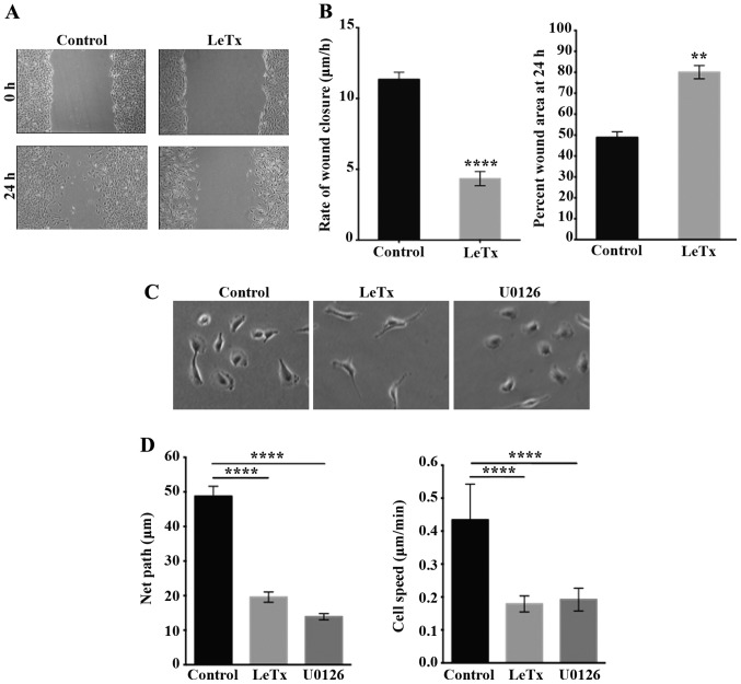
Recombinant anthrax lethal toxin inhibits 2D motility. (A) Cells treated with the recombinant anthrax lethal toxin for 2 h were grown in a monolayer then wounded and left to recover the wound then imaged at the same frame after 24 h (lower micrographs). Scale bar, 50 μm. (B) Quantitation for (A). Left panel, wound widths were measured at 12 different points for each wound, and the average rate of wound closure for the cells was calculated in μm/h. Right panel, percentages of wound area at 24 h following wounding. Data are the mean ± SEM from three wounds closure assays from three independent experiments. ****P<0.0001 indicates a statistically significant difference; **P<0.002 indicates statistically significant differences. (C) Representative micrographs of cells treated with either recombinant anthrax lethal toxin or the MEK inhibitor U0126 and allowed to undergo random motility in serum. Scale bar, 50 μm. (D) The net paths (left panel) or the cell speed of projected 120 frames from 2 h long time lapse movies were quantitated and expressed in μm and μm/min, respectively. Data are the mean ± SEM from 15 movies. ****P<0.0001 indicates statistically significant differences.

