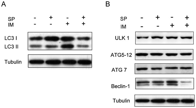
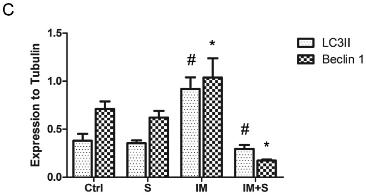
Figure 1.
Spautin-1 inhibits IM-induced autophagy in K562 cells. After treatment with or without IM (250 nM) for 12 h, spautin-1 (10 μM) or DMSO (0.1%) was added to K562 medium for further 36 h. (A) Autophagy was denoted by the switch of LC3I to LC3II, which was detected by western blotting (WB). (B) Other autophagy factors such as Beclin-1, ULK1, ATG 5 and ATG 7 were also detected by WB. (C) The bar chart demonstrates the ratio of LC3II and Beclin-1 proteins to tubulin by densitometry. Data are expressed as mean ± SD (#,*P≤0.05 between groups).


