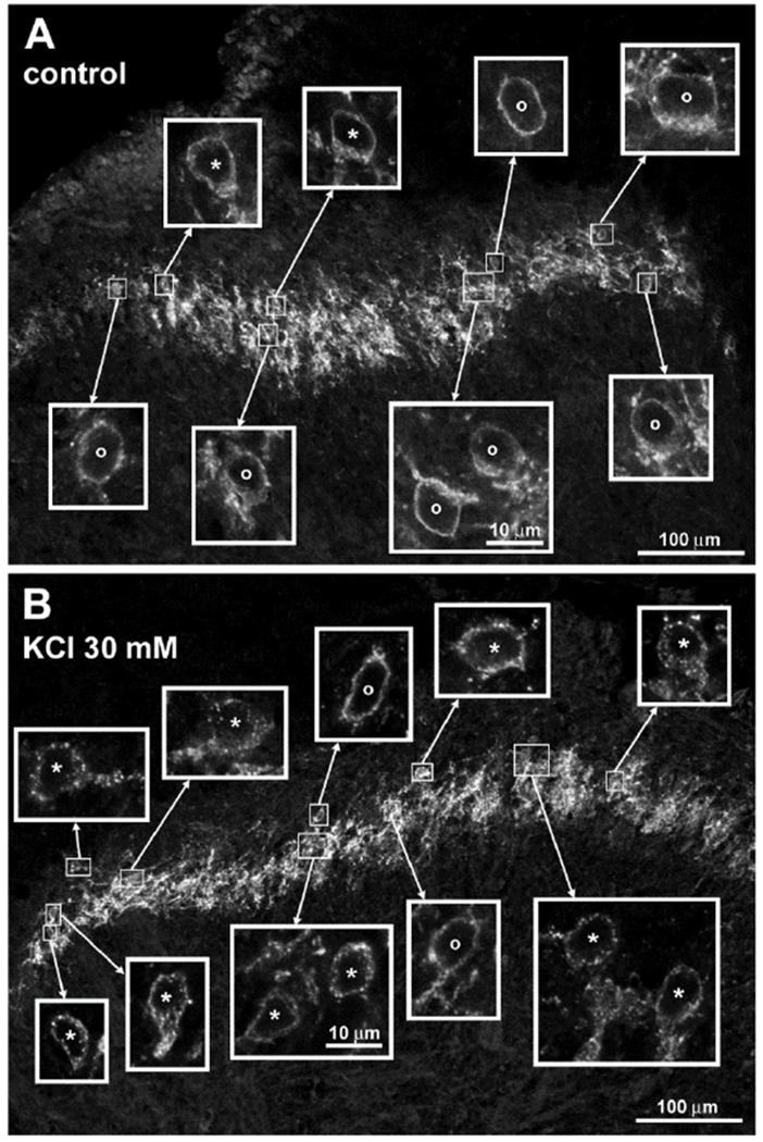
Figure 1 -. Y1 receptor immunoreactivity in the rat spinal cord and its internalization by endogenously released NPY.

A. Untreated rat spinal cord slice. B. Spinal cord slice incubated for 2 min with 30 mM KCl and then for 8 min in aCSF. In histological sections from the slices the Y1 receptor antibody labeled cells and fibers in lamina II. The fluorophore was Alexa Fluor 546. Main panels are images taken with a 20x objective and a zoom of 0.6, 10 optical sections (A) and 9 optical sections (B) separated 0.96 μm. Insets are images taken with a 63x objective and a zoom of 2.0, 4 optical sections separated 0.42 μm. Cells labeled ‘o’ and ‘*’ were considered without and with Y1 receptor internalization, respectively. All insets are at the same scale.
