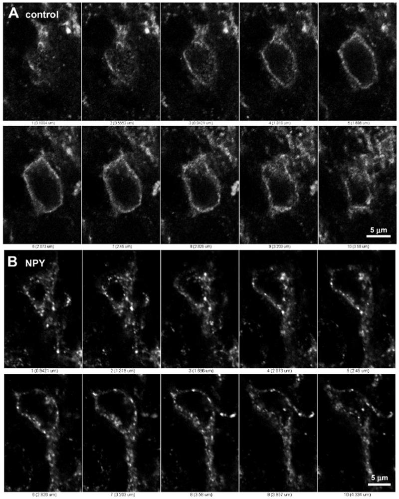
Figure 2 -. Lamina II neurons without and with Y1 receptor internalization.

Series of confocal optical sections separated 0.38 μm taken with a 63x objective through two lamina II neurons labeled with the Y1 receptor antibody and Alexa Fluor 488 secondary antibody. A: Neuron from a control spinal cord slice; the Y1 receptor is located at the cell surface and no endosomes are observed. B: Neuron from a spinal cord slice incubated with 1 μM NPY for 10 min; the Y1 receptor is located in endosomes inside the cytoplasm and one dendrite, but not in the nucleus.
