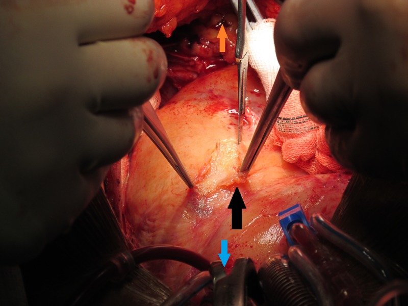
Figure 3.

Intraoperative image demonstrating the well-formed pericardial adhesions over the left ventricle, shown by the black arrow. The blue arrow is pointing towards the head of the patient and the orange arrow is pointing towards the patient’s feet.
