Fig. 3.

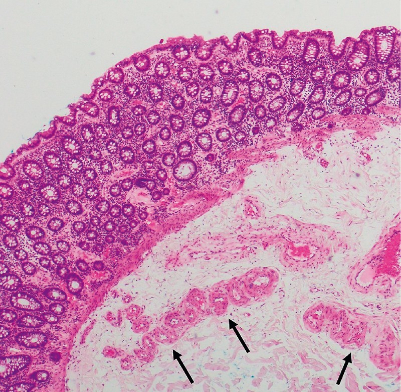
Histopathology from resected colonic angioectasia. Hematoxylin & eosin stain × 40. Dilated, clustered vessels in mucosa and submucosa, with tortuous feeding vessel at the resection base (marked with black arrows).

Histopathology from resected colonic angioectasia. Hematoxylin & eosin stain × 40. Dilated, clustered vessels in mucosa and submucosa, with tortuous feeding vessel at the resection base (marked with black arrows).