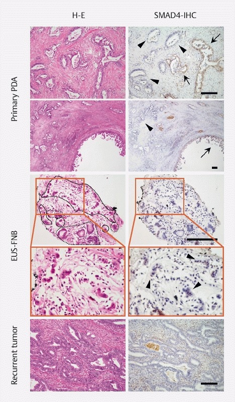
Fig. 3.

Immunostaining for SMAD4 in the surgically resected and endoscopic ultrasound-fine needle biopsy (EUS-FNB) specimens. SMAD4 staining was reduced (arrows) or absent (arrowheads) in the surgically resected specimen (upper panel). Loss of SMAD4 expression was noted in the majority of the tumor cells in the EUS-FNB specimens (middle) as well as in the recurrent gastric wall tumor (bottom). Areas marked with a solid line in the middle panel indicate atypical cells suggestive of adenocarcinoma, while dashed lines denote gastric foveolar epithelia. Scale bars: 200 µm.
