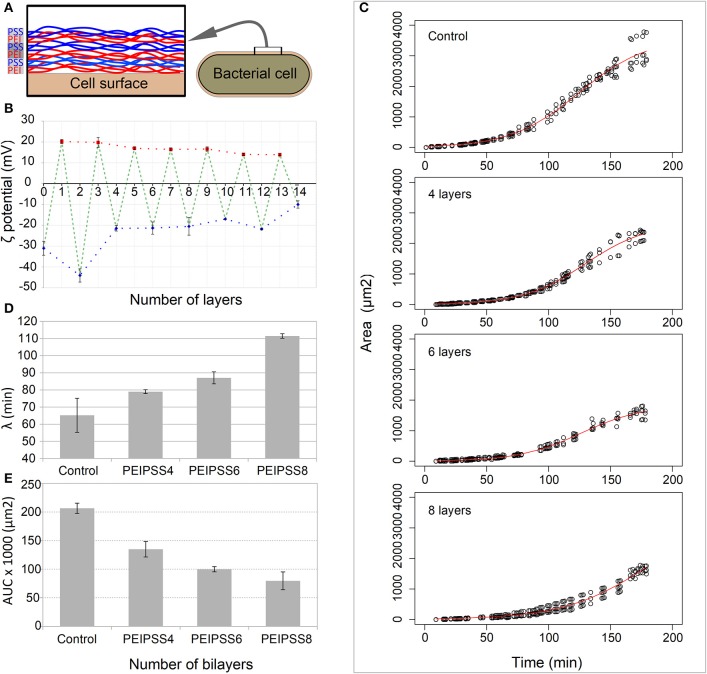
Figure 2.
Effects of different number of layers of growth of bacterial cells on a single cell level observed by time-lapse confocal microscopy. (A) Bacterial cells were coated with bilayers of polyelectrolytes always ending with a negative one. (B) The deposition of layers on bacterial cells was determined by the measurement of the zeta potential. (C) The delay of lag phase is correlated with the increasing numbers of layers, and was evaluated by determining the time point of the end of (D) the lag phase, the lambda. (E) The decreased area under the curve for more than 4 layers resulted from the curve shift toward the right.

