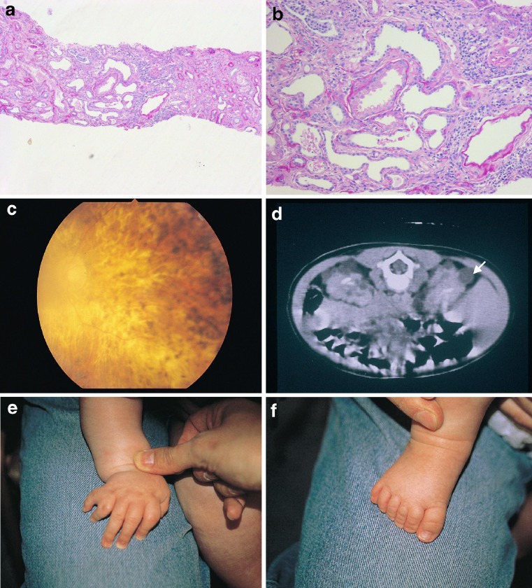
Fig. 3.
Examples of renal histopathology in BBS patients. a, b Low and high power micrographs showing tubular dilatation in a biopsy from a BBS patient’s kidney (Histopathological sections courtesy of Dr. Neil Sebire, Great Ormond Street Hospital). c Fundoscopy showing retinitis pigmentosa with cataract. d Abdominal CT scan documenting cystic kidneys (arrowed). e, f Post-axial polydactyly in a hand and foot from the same child with BBS

