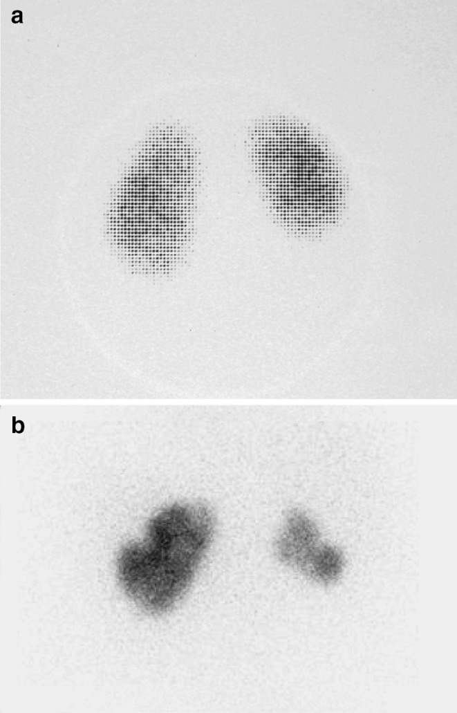
Fig. 1.

a DMSA scan of a 2-year-old girl with persistent, bilateral grade II VUR and dysfunctional voiding 6 months after the first documented febrile UTI. A smaller size of right kidney is demonstrated, compared to the left kidney with focal and generalized reduction in radiotracer uptake in the poles and indentation of the renal contour. The left kidney also presents a lack of homogeneity in DMSA uptake, mainly in the lower pole. b A DMSA scan 9 years later, after stopping the follow-up and antibiotic prophylaxis on the family’s own initiative and after breakthrough UTIs. The right kidney demonstrates further reduction of the size, and new scars are seen in both kidneys
