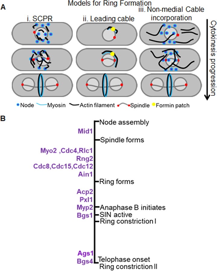
Figure 1. Early cytokinetic events.
A. Models for actomyosin ring assembly. i. The search-capture-pull-release model- the formin Cdc12 when recruited to the nodes nucleates actin while the type 2 myosin binds actin and pulls on the filaments to coalesce into a ring. ii. The Leading cable model- A patch of formin, Cdc12, nucleates actin cables that extends along the periphery of the division site to eventually form a ring. iii. Non-medial cable incorporation model. The formins Cdc12 and For3 nucleate actin to form non-medial cables that are then incorporated into the cells medial region to for a ring. B. Timeline of key proteins recruited to the division site leading to ring constriction with reference to different cytokinetic steps and mitotic progression. Biphasic ring constriction is depicted as Ring constriction I that occurs during anaphase B after Bgs1 recruitment and is slow and as Ring constriction II that occurs during telophase after recruitment of Ags1 and Bgs4 and is accelerated.

