Figure-2:
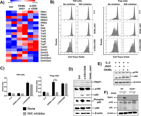
A) CD4+ T-cells from Foxp3.GFP mice were treated with OX40L-JAG1 or α-CD3/CD28 for 3 days. CD4+Foxp3-GFP- Teff cells and CD4+Foxp3+GFP+ Tregs were sorted and subjected to microarray analysis. Unstimulated fresh Teff cells and Tregs used as a control. Heat map shows differential mRNA expression of TRAFs, NF-kB and DNMT signaling related genes between control, OX40L-JAG1 and α-CD3/CD28 treated Tregs. B) CD4+CD25- Tconv cells and CD4+CD25+ Treg cells were pre-treated with indicated NIK inhibitor (10μM/ml) for 2h and co-treated with IL-2 (25U/ml) and OX40L (5 μg/ml) + JAG1 (1 μg/ml) and αCD3+ αCD28 monoclonal antibodies in the presence of IL-2 for 4 days. Cell proliferation was measured by cell trace violet dilution assay. C) Bar graphs show division index calculated from cell trace violet dilution in each culture conditions (Values represent Mean ± SEM, n=3, ****p<0.001). D) Western blots show TRAF1 expression, p100 to p52 processing and total and phospho p65 levels in OX40L-JAG1 treated Tregs compared to fresh and α-CD3/CD28 treated Tregs after 24hr. E) Western blots show p100 to p52 processing in Tregs treated with IL-2 alone, JAG1 alone, OX40L-alone and OX40L+JAG1+IL-2. F) Western blots show TRAF1 and p100 to p52 processing in WT and OX40−/− Tregs treated with or without OX40L.

