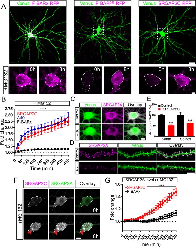
Figure 2.
Proteasome degradation of SRGAP2 proteins. (A) Expression of RFP-tagged F-BARx, F-BARΔ49, or SRGAP2C in mouse cortical pyramidal neurons cultured for 18–21 days in vitro (DIV). Low levels of expression were observed for F-BARΔ49 and SRGAP2C, while addition of the proteasome inhibitor MG-132 resulted in a strong increase after 8 h of treatment. F-BARx is highly expressed throughout the experiment. Scale bar top panels: 25 µm, bottom panels: 5 µm. (B) Quantification of fluorescence intensity after MG-132 treatment. nF-BARx = 31 neurons, nF-BARΔ49 = 50 neurons, nSRGAP2C = 47 neurons; multiple t tests with Holm-Sidak correction for multiple comparisons (α = 0.05); ****p < 0.0001; mean ± SEM. (C–E) Co-expression of SRGAP2A and SRGAP2C in cultured mouse cortical neurons. SRGAP2A levels are reduced when co-expressed with SRGAP2C in both soma and dendritic spines. Scale bars: 5 µm. (E) Quantification of SRGAP2A fluorescence intensity. Soma: nControl = 110, nSRGAP2C = 68; Spines: nControl = 969, nSRGAP2C = 1259; Mann-Whitney test; ***p < 0.001; mean ± SEM. (F) Treatment of cultured mouse cortical neurons expressing both SRGAP2A-GFP and SRGAP2C-RFP with MG-132. Levels for both SRGAP2A and SRGAP2C increased upon treatment, and co-localization of SRGAP2A and SRGAP2C protein clusters were observed (red arrow). Scale bar: 10 µm. (G) Quantification of SRGAP2A fluorescent intensity when co-expressed with either F-BARx or SRGAP2C in the presence of MG-132. nF-BAR = 60 neurons, nSRGAP2C = 59 neurons; multiple t tests with Holm-Sidak correction for multiple comparisons (α = 0.05); ***p < 0.001; mean ± SEM.

