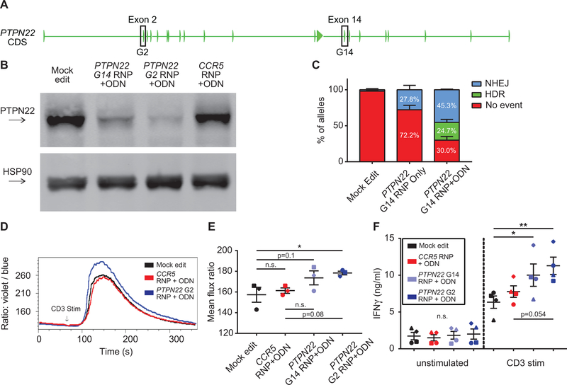
Figure 2. Disruption of PTPN22 in CD4+ cells results in increased TCR-triggered calcium flux.
(A) PTPN22 coding exons with targeted exons highlighted. (B) Representative western blot of PTPN22 expression in mock, PTPN22 G14, PTPN22 G2, and CCR5 edited CD4+ T cells from the same human donor. Cells were expanded 7 days post-editing and rested 24 hours in cytokine free media, as in Fig. 1A, prior to lysis. (C) ddPCR analysis of editing frequencies in unedited and PTPN22 G14 edited cells +/− stop codon containing ssODN (bars represent mean +/− SEM, n=4 independent human donors, percentages reflect summary data). (D) Representative TCR-induced calcium flux of human CD4+ T cells generated, expanded, and rested as in Fig.1A. Cells were stained with indo-1 AM, monitored for baseline, then stimulated with anti-CD3 (arrow). (E) Summary of mean flux ratios for data generated as in (D) (graphs lines and error bars represent mean +/− SEM, n=3, matched one-way ANOVA with Tukey’s correction). (F) IFNγ ELISA using supernatants from mock or edited cells +/− stimulation with plate bound anti-CD3 for 48 hours. RNP – ribonucleoprotein, ODN or ssODN - single stranded oligo-deoxynucleotide, NHEJ - non-homologous end joining, HDR - homology directed repair. Shapes in summary plots correspond to individual donors. All data is from at least 2 independent experiments. * p<0.05, ** p<0.01, *** p<0.001, **** p<0.0001.

