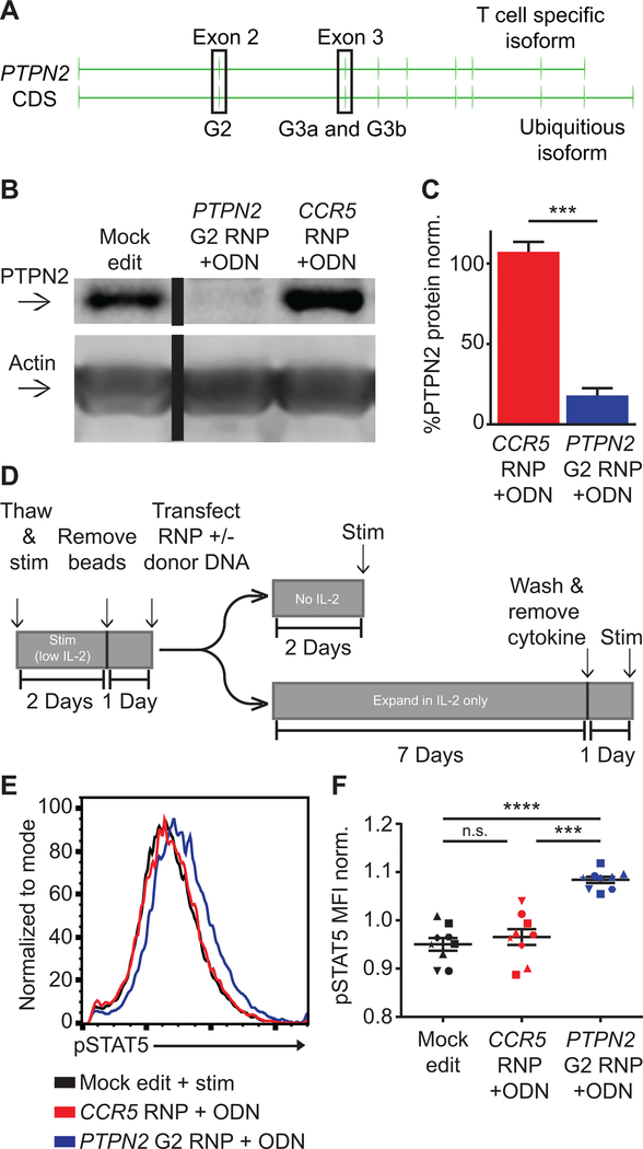
Figure 5. PTPN2 disruption in CD4+ cells promotes increased IL-2 signaling.
(A) PTPN2 gene showing structure and exons used for alternative isoform expression and location of guide RNA target sites (in highlighted exons). (B) Representative western blot of PTPN2 expression in mock, PTPN2, and CCR5 edited CD4+ T cells from the same human donor. Cells were expanded 7 days post-editing and rested 24 hours in cytokine free media, as in Fig. 1A, prior to lysis. Lanes were run on the same gel but were non-contiguous. (C) Quantified PTPN2 protein expression relative to actin and normalized to mock edited values from the same T cell donor (bars represent mean +/− SEM, n=6 independent human donors, paired t test). (D) Work-flow used to produce and assay PTPN2 edited CD4+ T cells and corresponding controls with or without IL-2 supplemented media. (E-F) Human CD4+ T cells edited as in (A) and rested for 2 days in cytokine free media, were stimulated with IL-2 for 20 minutes. (E) Representative histogram overlay of pSTAT5 in mock, PTPN2, and CCR5 edited cells from the same donor. (F) Summary flow data of median pSTAT5 expression post IL-2 stimulation (graph lines and error bars represent mean +/− SEM, n=9 human samples (7 independent donors plus 2 repeat donors, repeat donors were run in separate experiments), matched one-way ANOVA with Tukey’s correction). RNP - ribonucleoprotein, ODN or ssODN - single stranded oligo-deoxynucleotide. Shapes in summary plots correspond to individual donors. All data is from 2 or 3 independent experiments. * p<0.05, ** p<0.01, *** p<0.001, **** p<0.0001.

