Figure 1.
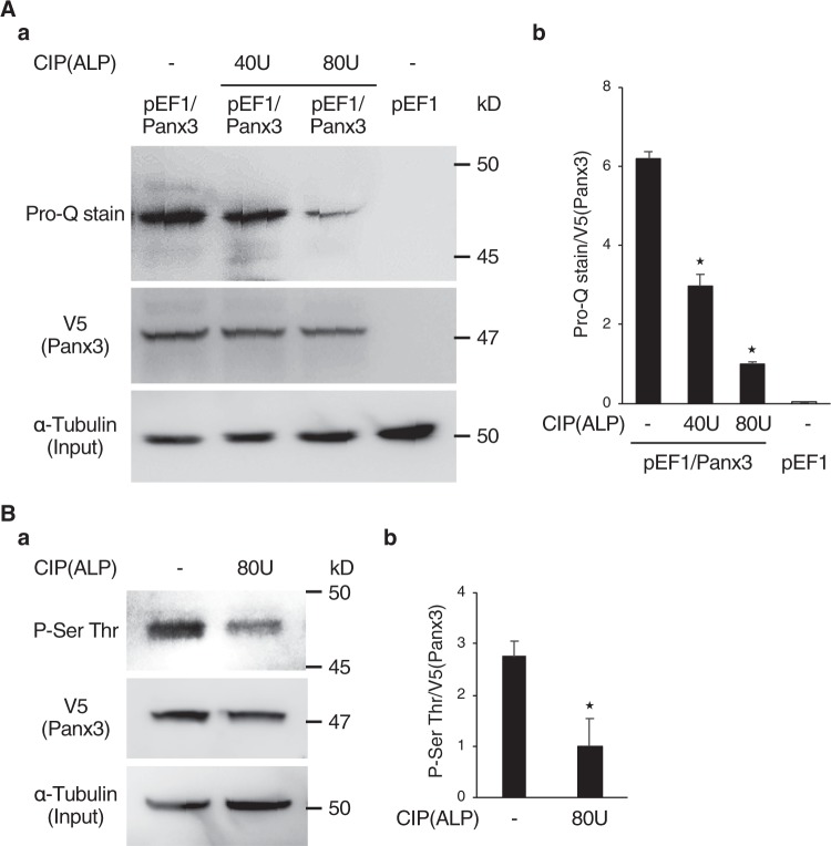
Panx3 is phosphorylated. (A,a) A phosphorylation band was revealed from pEF1/Panx3 cell lysates by Pro-Q Diamond phosphoprotein gel staining. Control (pEF1) cells and C2C12 cells transiently transfected with Panx3 (pEF1/Panx3) were cultured for 2 days in DMEM containing 10% FBS. Immunoprecipitated cell lysates were loaded onto NuPAGE Bis-Tris gels. After electrophoresis, the gels were fixed and stained with Pro-Q stain. Lysates were incubated with CIP (ALP:40 or 80 U) for 45 min at 30 °C before loading the gel. Lysates were analyzed by western blotting with a α-tubulin as a control, to confirm loading of the same amount of protein (bottom). (b) Quantification of the ratios of Pro-Q stained band/V5. (B,a) Immunoprecipitation assays with cell lysates from pEF1/Panx3 transiently transfected cells showed a phosphorylation band of serine and threonine which was reduced by CIP. The Panx3 protein overexpressed in C2C12 cells was precipitated with anti-V5 antibody and analyzed by immunoblotting with Phospho-Serine and Threonine (P-Ser Thr) antibody. Precipitated lysates were treated with CIP (80U) before loading. (b) Quantification of the ratios of P-Ser Thr/V5. *P < 0.01. Error bars represent the mean ± SD; n = 3. Pro-Q stain and Western blot were perfomed by at least three independent experiments for each experimental group. Full blot is shown in the Supplemental Information (Full Original Blots-I).

