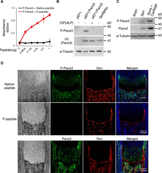
Figure 3.
Generation of P-Panx3 specific antibody and its localization in the tibial growth plate of newborn mice. (A) Specificity evaluation of phosphorylated Panx3 at Ser68 antibody (P-Panx3) against antigen-peptide (P-peptide) or control (Native peptide) by ELISA. ELISA was performed in dishes coated with serial dilutions of P-Panx3 peptide. Error bars represent the mean ± SD; n = 6. (B) Western blots, using P-Panx3 antibody and lysates from C2C12 cells stably transfected with pEF1/Panx3 and Ser68Ala. Protein was isolated after 1 day of culture in DMEM. For dephosphorylation, protein from pEF1/Panx3 cells was treated CIP (80U) before loading. V5 antibody was used for detection of Panx3 expression. (C) Western blots using P-Panx3 and Panx3 antibodies and tissue lysates isolated from brain, skin and bone + cartilage of newborn mice. (D) Immunohistochemistry of newborn mouse growth plates. Image observed under light microscopy (black & white, left vertical lane), P-Panx3 (green, upper and middle lane), Panx3 (green, bottom lane), Ocn (red), Hoechst nuclear staining (blue) and merged image (right vertical lane) The middle lane is staining images of P-Panx3 in the presence of its antigen peptide (P-peptide). Western blot was perfomed by at least three independent experiments. Full blot is shown in the Supplemental Information (Full Original Blots-III).

