Figure 4.
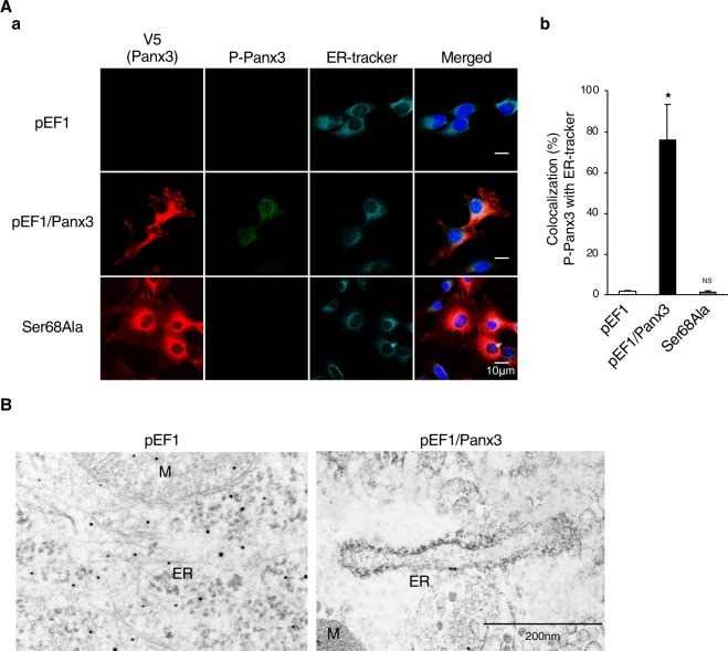
Phosphorylated Panx3 at Ser68 is localized on the ER membrane. (A,a) Cellular localization of P-Panx3 in cells stably transfected with pEF1, pEF1/Panx3, or Ser68Ala. Fluorescent confocal images showed Panx3 (red), P-Panx3 (green), ER-tracker (cyan), and Hoechst nuclear staining (blue). (b) Measurements show a percentage of colocalization between P-Panx3 with ER-tracker. (B) Immuno-TEM demonstrates P-Panx3 localization on the ER membrane in pEF1/Panx3 transfected cells (right panel). M: mitochondria, ER: endoplasmic reticulum. The micrographs shown are representative of 15–20 cells obtained from three independent experiments. *P < 0.01. NS, nonsignificant. Error bars represent the mean ± SD; n = 12.

