Figure 5.
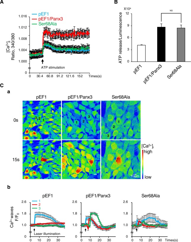
Phosphorylation at Ser68 regulates the functioning of the Panx3 ER Ca2+ channel, but not the hemichannel or gap junction. (A) ER Ca2+ channel activities in cells stably transfected with pEF1, pEF1/Panx3 or Ser68Ala. The increase in intracellular Ca2+ released from the ER by ATP stimulation (arrow) was measured. Average traces of at least four different experiments are shown with the solid lines. The arrow indicate the time of ATP stimulation. (B) Hemichannel activities in each stable cell line. ATP release into the extracellular space was measured for 2 min. NS, nonsignificant. (C) Gap junction activities in each stable cell line. (a) Representative images of the Fluo-4 fluorescence before stimulation (upper) and 15 s after the stimulation (bottom) and the fluorescence intensity traces of laser stimulated single cell (1), one (2) and two cell (3) distant to stimulated single cell (b). Average traces of at least three different experiments are shown with the solid lines. The arrow indicate the time of ATP stimulation. The Ca2+ wave was measured in cells loaded with Fluo-4 and NP-EGTA (caged Ca2+) by starting uncaging in a single cell using laser illumination. The Ca2+ wave propagation was measured at 30 s after the illumination. The images shown are representative of at least three different experiments.

