Figure 1.

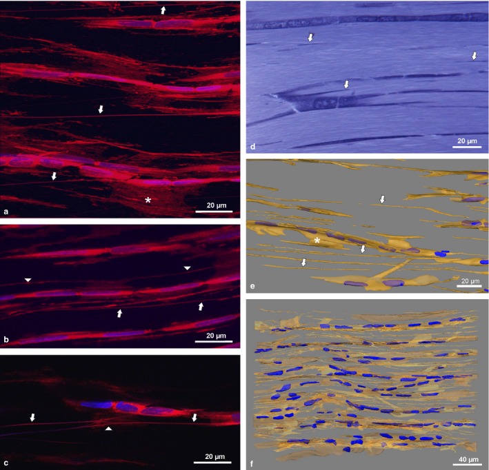
Confocal images of whole mount adult rat tail tendon fascicles (RTTfs) stained with Deep Red and DAPI nuclear stain (a–c) showing sheet‐like processes (*) and NTs (arrows) connecting tenocytes. Arrowheads depict shorter NTs connecting cell sheets of tenocytes in the same cell row (b) and links or potential branching of NTs (c), respectively. Semithin section of RTTfs of a 9‐week‐old rat after toluidine blue staining (d) and 3‐D reconstruction (e,f) showing NTs (arrows). For detailed display of the 3‐D reconstruction see Video S1.
