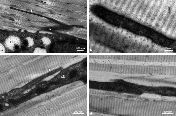
Figure 2.

TEM micrographs of adult rat tendons showing cell bodies (cb) of tenocytes with a lateral sheet‐like process (*) and a longitudinally directed NT (arrow) originating from the cell sheet (a) and the cell body (c); detail of NT (b) and closed NT end‐to‐end contact (d).
