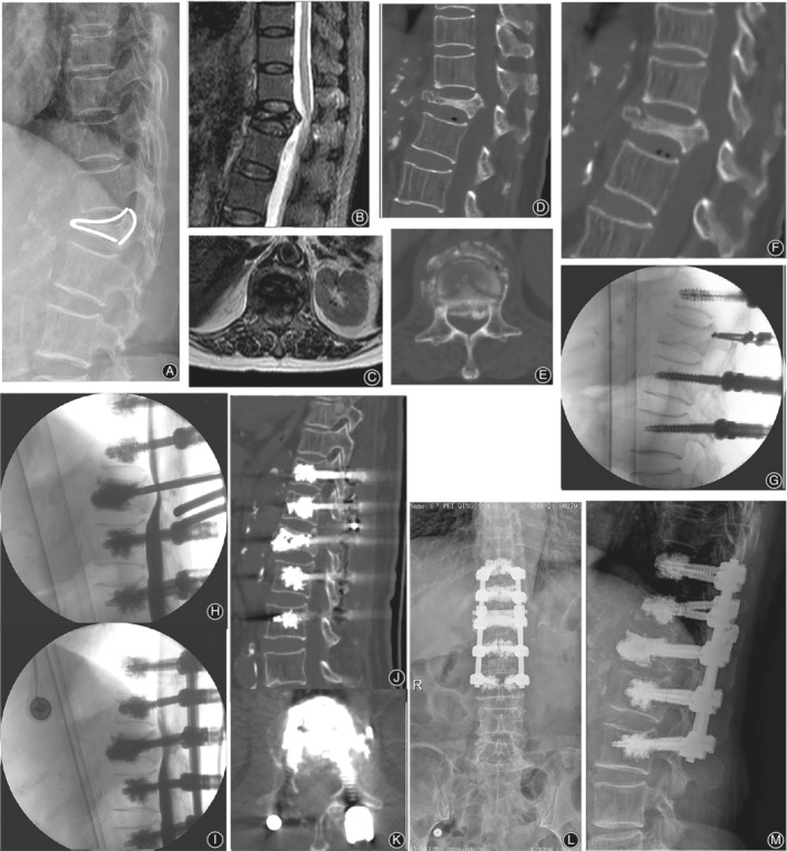
Figure 4.

Representative case of type 2 OVF. A 76‐year‐old female underwent VP and limited laminectomy at L1, combined with posterolateral bone fusion and instrumentation from T11 to L3 with augmented pedicle screw fixation. Preoperative lateral radiographs (A) and T2‐weighted MRI scans (B,C) show that significant T12 collapse with dural compression. Barely any height restoration or canal decompression is shown on extension CT (D, E). Intraoperative fluoroscopy (G) shows restoration of the vertebral height without dural decompression on myelography (H, I). Postoperative CT scans (J, K) and lateral radiographs (L, I).
