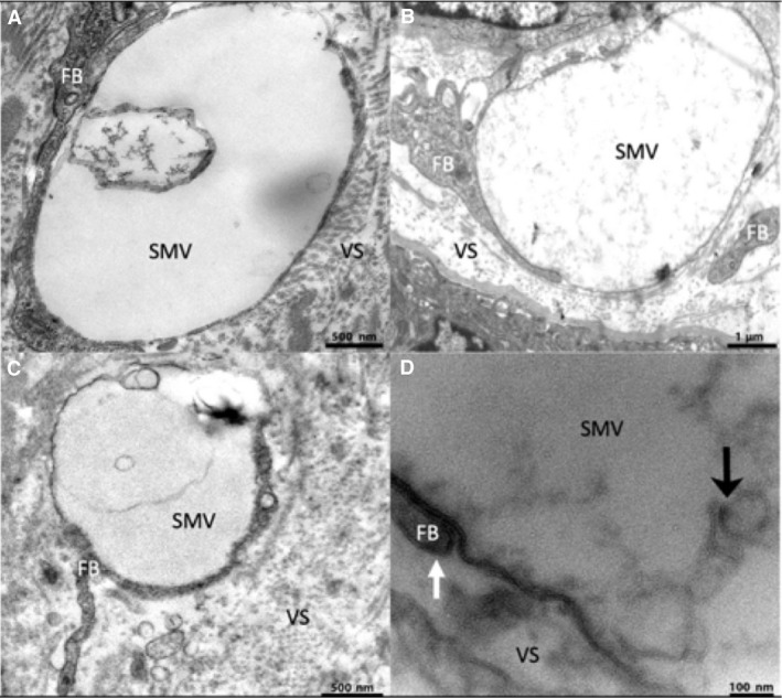
Figure 2.

Transmission electron microscopy images of extracellular stromal macrovesicles showing contact with stellate processes and internal contents. (A) A stromal extracellular vesicle containing a membranous component in its cavity. (B) An extracellular vesicle containing diffuse material. (C) An extracellular vesicle containing a membranous component. (D) A higher magnification image showing the lipid bilayer of the vesicle membrane and an adjacent fibroblast process (white arrow) as well the membranous components (black arrow) inside the cavity of an extracellular stromal macrovesicle. FB, fibroblast‐like stellate cell; SMV, stromal macrovesicle; VS, villous stroma.
