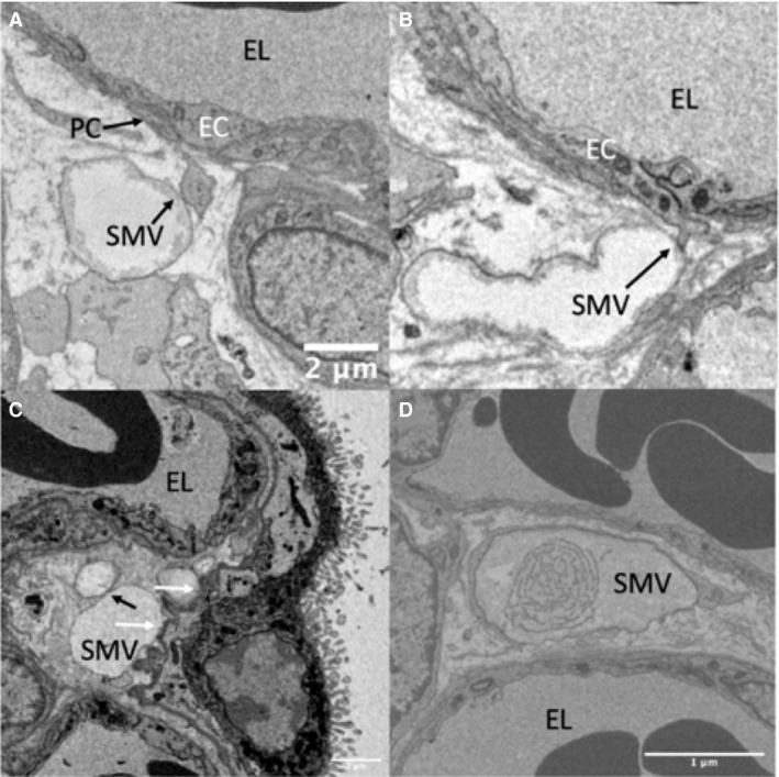
Figure 3.

Serial block‐face scanning electron microscopy images of stromal macrovesicles making contact with structures other than stellate cells. (A) A pericyte process touching a stromal microvesicle. (B) An endothelial cell process touching a stromal macrovesicle (C) Shows stromal macrovesicles lying adjacent to the syncytiotrophoblast basal lamina (contact points indicated by a white arrow) and also adjacent to a separate vesicle (contact point indicated by a black arrow; these vesicles were shown to be distinct structures by inspecting serial sections). (D) An example of a vesicle whose contents had a higher degree of structure than was typically observed. EC, endothelial cell; PC, pericyte; SMV, macrovesicle.
