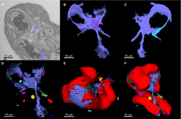
Figure 4.

Three‐dimensional reconstruction of a fibroblast‐like stellate cell and its relationship with capillaries and stromal extracellular macrovesicles. (A) Serial block‐face scanning electron microscopy image of a placental villus showing the segmented processes of a fibroblast‐like stellate cell emerging from the slice (blue). Both of the blue segmented regions are part of the same cell. (B) Segmented three‐dimensional fibroblast structure. (C) Regions on the stellate cell where there were interactions with other stellate cells are shown in light blue. (D) Fibroblast interactions with extracellular macrovesicles in the villous stroma (see Fig. S1 for an interactive 3D model). (E) The relationship between the fetal capillary and the fibroblast. (F) Association of the stellate cell with the fetal capillary and the extracellular macrovesicles.
