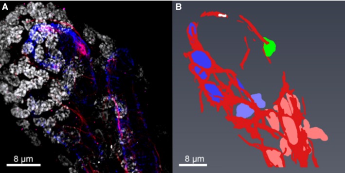
Figure 5.

Wholemount confocal imaging of networks of fibroblast‐like stellate cells. (A) A projection of a confocal stack showing fibroblast‐like stellate cell networks stained with SLC22A11 (red), pericytes surrounding fetal capillaries stained with α‐SMA (blue), and the nuclei stained with DAPI (white). (B) Segmentation of the fibroblast‐like stellate cell networks shown in (A). The fibroblast‐like stellate cell processes are shown in red and their nuclei are shown in four different colours [blue, light blue, green (isolated cell) and pink], demonstrating three different networks.
