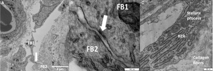
Figure 6.

Electron microscopy images of placental stromal stellate cells. (A) A single serial block‐face scanning electron microscopy image showing the networks of stellate processes within the stroma with the white arrow indicating the junctional complex between two stellate cells (FB, fibroblast‐like stellate cell). (B) Transmission electron microscopy (TEM) image of two adjacent stellate cells connected with an adherens junction (white arrow). The two stellate cells are marked FB1 and FB2. (C) TEM image of a fibroblast process containing cellular machinery. RER, rough endoplasmic reticulum.
