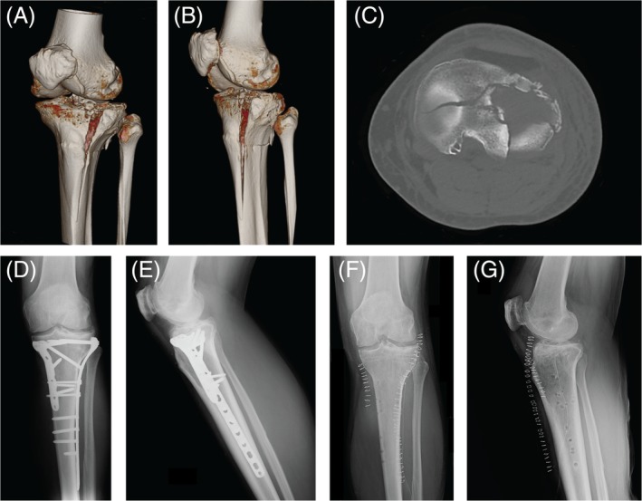
Figure 1.

Case 1: A 56‐year‐old man with an AO 41‐C3 fracture treated by open reduction internal fixation (ORIF) technique. (A, B) Preoperative 3D‐CT scan reconstruction images; (C) CT axial image; (D, E) Anteroposterior and lateral radiographic images 6 months after surgery; (F, G) Anteroposterior and lateral radiographic images after removal of implants 35 months after surgery.
