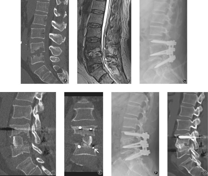
Figure 1.

Preoperative CT (A), MRI (B) of a 63‐year‐old man suffering from L4/ 5 spondylodiscitis with partial destruction of the vertebral bodies. Immediately post‐surgery X‐ray (C) and CT scan (D, E). Both the L5 pedicle screws were placed below the cortical vertebrae (E, white arrow). One year after surgery, X‐ray (F) and CT (G) scan showed a solid fusion of the bone graft and vertebral body interface.
