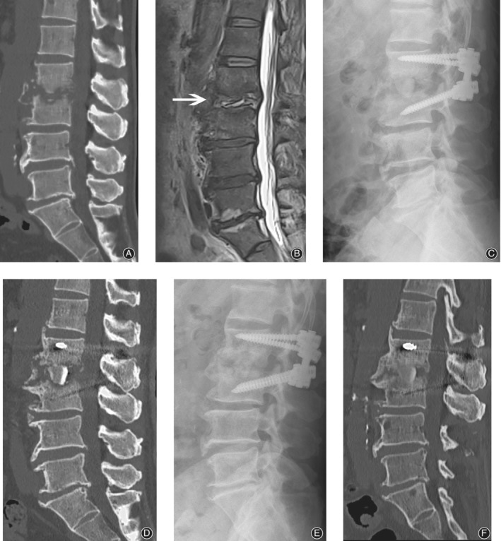
Figure 2.

Sixty‐two‐year‐old woman, whose chief complaint was back pain of more than 3 months. Pre‐operative CT (A) and MRI (B) images revealed L2– 3 intra‐vertebral space infection with both upper and lower endplate destruction. Debridement and reconstruction underwent via oblique lateral interbody fusion (OLIF) corridor and posterior approach. A massive structure bone graft was seen in the immediately postoperative film (C) and CT scan (D). Post‐surgery 1 year, both the film (E) and CT scan (F) showed perfect fusion between bone graft and vertebrae interface. White arrow indicates the index level.
