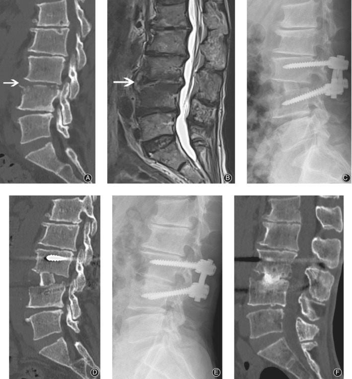
Figure 3.

Fifty two‐year old man, with back pain for 2 months without fever. Pre‐operative CT scan (A) showed superior endplate destruction of L4. Pre‐operative MRI (B) shows high T2 signal in disc space and low T2 signal in both upper and lower endplate. Surgery was performed via as mentioned method. A massive structure bone graft was seen in the immediately postoperative film (C) and CT scan (D). One year after surgery, fusion was achieved between the interface in both film (E) and CT (F) scan. White arrow indicates the index level.
