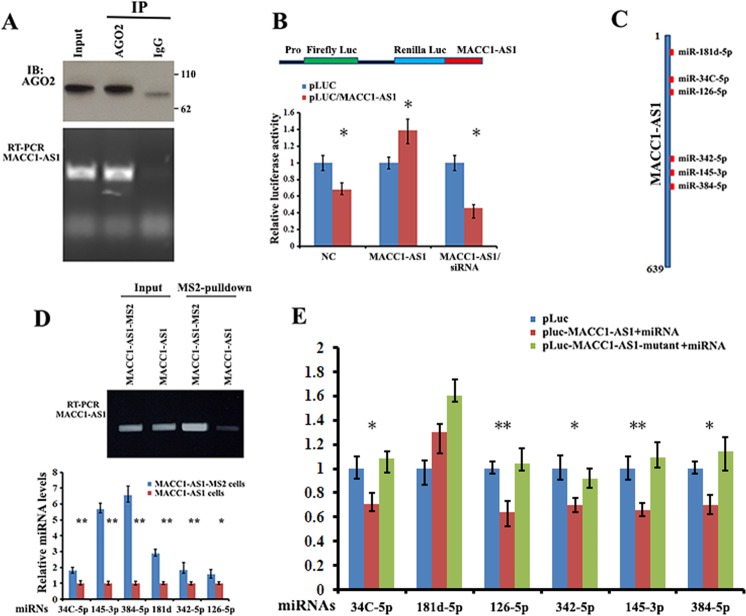
Fig. 3. MACC1-AS1 serves as a platform for Ago2 and miRNA binding.
a Upper: immunoprecipitation (IP) of Ago2 protein from extracts of HEK-293T cells. Lower: levels of MACC1-AS1 co-precipitated with Ago2 protein were analyzed by RT-PCR. b Upper: schematic of a luciferase reporter in which the entire MACC1-AS1 sequence was fused into the 3′ region of the Renilla luciferase gene of the psiCHECK-2 construct (denoted pLuc-MACC1-AS1). Lower: HEK-293T cells were transfected with pLuc (control) or pLuc-MACC1-AS1, or pLuc-MACC1-AS1 in combination with either the MACC1-AS1 expressing vector or siRNA against MACC1-AS1. Luciferase activities were determined after 36 h transfection using a dual-luciferase assay system. Renilla luciferase activity was normalized to the activity of firefly luciferase. *P < 0.05 as determined by Student’s t test. c Schematic representation of the potential binding sites for individual miRNAs within MACC1-AS1 RNA. d Upper: RT-PCR and agarose gel electrophoresis indicate that MACC1-AS1-MS2 was pulled down by MBP-MCP-conjugated amylose resin. Lower: individual miRNAs were detected by RT-qPCR in the precipitates of MACC1-AS1. **P < 0.01 as determined by Student’s t test. e HEK-293T cells were co-transfected with pLuc, wild-type or mutant MACC1-AS1 reporters with the corresponding miRNA mimics. Luciferase activity was determined by the dual luciferase reporter system. Activity of Renilla luciferase was normalized to the activity of firefly luciferase. **P < 0.01, *P < 0.05 as determined by one-way ANOVA followed by Tukey’s multiple comparison tests.

